Chondroid Matrix X Ray

Pin By Phuong Hoai On Msk In 2020 Radiology Imaging Medical Medicine

Pin By Tnfri On Radiologi In 2020 Radiology Imaging Human Anatomy And Physiology Radiology

Pin By Phuong Hoai On Musculoskeletal Radiology Bone Diseases Pathology

Aneurysmal Bone Cyst Cysts Medical News First Aid Tips

Pin By محمد البدوي On Arthritis In 2020 Subtle Lockscreen Movie Posters
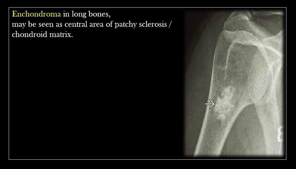
Pin By Raghav Tiwari On Msk In 2020 Central Area Arthritis Patchy
They are most commonly found in older patients within the long bones and can arise de novo or secondary from an existing benign cartilaginous neoplasm.
Chondroid matrix x ray. Authors experience and a. Bachoura a rice is lubahn ar lubahn jd. The management and surgical intervention timing of enchondromas. On imaging these tumors have ring and arc chondroid matrix mineralization with aggressive features such as lytic pattern deep endosteal scalloping and soft tissue extension.
To present the different techniques. In general well formed rings and arcs are seen in lower grade tumors see chondrosarcoma. Zhou x zhao b keshav p chen x gao w yan h. Although chondroid matrix is highly recognizable it can sometimes be difficult to distinguish from bone infarcts especially on x ray since both have well defined irregular margins with areas of fluid signal intensity and apparent calcification or ossification.
To illustrate the spectrum of chondroid matrix lesions. Plain x ray computed tomography ct magnetic resonance mri and pet ct available for the study of chondroid matrix lesions and their role in the diagnostic procedures. The surgical management of hand enchondroma without postcurettage void augmentation. They are lytic lesions that usually contain calcified chondroid matrix a rings and arcs pattern of calcification except in the phalanges.


Pin By Bassam Barca On Musculoskeletal Bone Diseases Sonography Pathology

Sclerotic Bone Tumors And Tumor Like Lesions Radiology Radiology Imaging Oral Pathology

Pin By Mauricio Zapata On Musculoskeletal Radiology Bone Diseases Pathology

Bonefixator Com Fractures And Healing 1 3 Fracture Healing Bone Healing Bone Fracture

Pin De Aaliaga En Msk Anatomia Humana Huesos Anatomia Humana Antropologia Forense

Pin By Phuong Hoai On Musculoskeletal Radiology Bone Diseases Pathology

